Fall 2019 (Volume 29, Number 3)
Rheumatology Art:
Foldscope Images
By Raman Joshi, MD, FRCPC
Download PDF
All these unique images were taken using a Foldscope, a paper origami microscope, attached to
an iPhone SE.
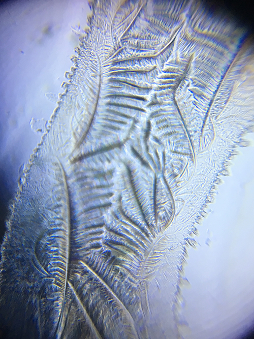
An image of avian-sourced hyaluronic acid. A drop of the hyaluronic
acid which was left over after injection was dried on a glass slide and
viewed with a foldscope.
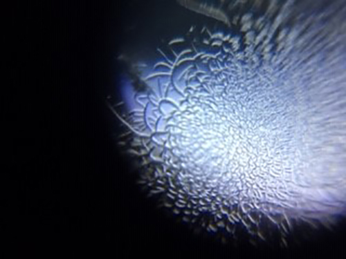
An image of etanercept, which had expired. A drop of fluid was dried on a
glass slide.
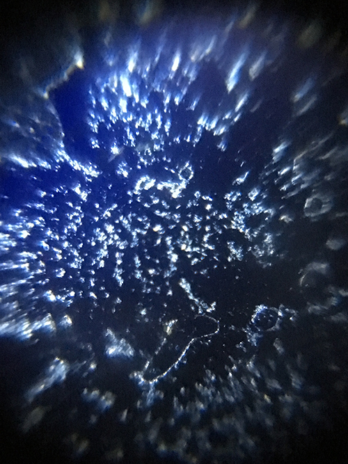
Triamcinolone hexacetonide
under polarized light.
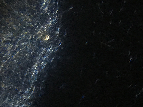
This image is of dried synovial fluid from a patient with acute inflammatory arthritis. The image
was taken using the iPhone and crossed polarizing plates to reveal the long, thin, negatively
birefringent crystals which were also seen by standard compensated polarizing microscopy and
are consistent with uric acid crystals.
|